Annotation Guidelines
This page gives an overview of the annotation and normalization scheme and process of the MedProcNER/ProcTEMIST corpus. More detailed information is available in the Annotation Guidelines, a 30+ pages long file that documents the corpus’s creation and annotation process. They are available on Zenodo.
The MedProcNER/ProcTEMIST guidelines were created by clinical experts at the same time as the DisTEMIST guidelines. After their definition, the guidelines were refined in several cycles of quality control and annotation consistency analysis before annotating the entire dataset, with a final agreement of… Additionally, once the manual annotation phase was finished, the corpus was thoroughly revised in a post-processing step to maximize consistency.
This page has two parts:
- Annotation
- Normalization
Annotation
The corpus includes only one label: PROCEDIMIENTO (clinical procedure). Despite this unicity, the label and corpus itself are very varied. Several kinds of procedures are annotated, including diagnostic, therapeutic, preventive and supportive procedures. Some specific examples are:
- Simple medical exploration and inspection methods (that require little or no instrumentation): These procedures involve the use of basic diagnostic techniques to examine a patient’s body for signs of illness or disease. Examples include listening to the lungs with a stethoscope (“auscultación pulmonar”, pulmonary auscultation), feeling the abdomen for abnormalities (“palpación abdominal”, abdominal palpation), or checking the patient’s neurological responses (“exploración neurológica”, neurological examination).
- Imaging tests: These procedures involve the use of advanced medical technology to produce images of the inside of the body, which can be used to diagnose and monitor various conditions. Examples include magnetic resonance imaging (MRI) of the brain (“RMN cerebral”), computed tomography (CT) of the chest with contrast (“TAC torácico con contraste”), or x-rays of the femur from the anterior-posterior (AP) view (“RX de fémur AP”).
- Other medical tests: These procedures involve the use of laboratory tests or other diagnostic tools to evaluate a patient’s health status or monitor their condition. Examples include a complete blood count (“hemograma”, hemogram), electrocardiogram (“electrocardiograma” or “ECG”) to measure heart function, or electroencephalogram (“electroencefalograma” or “EEG”) to measure brain activity.
- Administration of medications: These procedures involve the delivery of medications to treat or manage a patient’s medical condition. Examples include antibiotic therapy to treat bacterial infections (“antibioterapia”) or corticosteroids (“corticosteroides”) to reduce inflammation.
- Administration of blood, plasma, serums, bolus and continuous medication pumps: These procedures involve the delivery of fluids, nutrients, or medications directly into a patient’s bloodstream. Examples include a blood transfusion to replace lost blood (“transfusión de 2 concentrados de hematíes”) or fluid therapy to treat dehydration (“sueroterapia”).
- Simplified surgical treatments: These procedures involve minimally invasive or straightforward surgical procedures that can be performed relatively quickly and easily. Examples include removal of the prostate gland through an incision in the lower abdomen (“adenomectomía retropúbica”, retropubic adenomectomy) or placement of a testicular prosthesis (“se coloca prótesis testicular”).
- Surgical descriptions: These procedures involve detailed accounts of surgical procedures, including the steps involved, the instruments used, and any complications that may arise. Examples include “reconstructed with a chin graft and an arched titanium plate” (“se reconstruyó con injerto de mentón y placa de titanio arqueada”) or “the intercortical gaps were filled with cancellous bone obtained from the donor area” (“los gaps intercorticales se rellenaron de hueso esponjoso obtenido de la zona donante”).
As with many other clinical entities, detecting and annotating procedures in structured text can be quite complicated due to the use of descriptive language, abbreviations, multiple parts (i.e. anatomical entities or instruments) and even ambiguous wording.

Physical examination revealed a good general state of health, with normal abdomen and genitalia; rectal examination was compatible with grade I/IV prostate adenoma.
Urinalysis showed 4 red blood cells/field and 0-5 leukocytes/field; the rest of the sediment was normal.
Normal haemogram; biochemistry showed glycaemia of 169 mg/dl and triglycerides of 456 mg/dl; normal liver and kidney function. PSA of 1.16 ng/ml.
Urine cytology was repeatedly suspicious for malignancy.
Simple abdominal X-ray shows degenerative changes in the lumbar spine and vascular calcifications in both hypochondrium and pelvis.
Urological ultrasound revealed the existence of simple cortical cysts in the right kidney, bladder without alterations with good capacity and prostate weighing 30g.
IVU showed bilateral renal normofunctionalism, calcifications on the right renal silhouette and arrhotic ureters with addition images in the upper third...

With the diagnosis of stage T3bN0M0 renal neoformation with level II tumour thrombus, it was decided to intervene, together with the cardiac surgery service of our centre, performing sternotomy and left subcostal laparotomy and, after freeing the splenic angle of the colon by opening the retroperitoneum at the level of the mesentery, left radical nephrectomy with the nephrectomy specimen remaining anchored by the thrombosed renal vein. Subsequently, and under extracorporeal circulation with deep hypothermia and cerebral retroperfusion, the filter is removed by closing it and traction under fluoroscopic control from its insertion point at jugular level, with control by right auriculotomy of the possible dissemination to the lung of small thrombi during the removal manoeuvre, followed by resection of the renal vein and its ostium due to suspicion of infiltration and thrombectomy by traction to subsequently repair the defect with a Goretex® patch, for which reason the patient required anticoagulation with dicoumarinic drugs for 3 months until endothelisation of the synthetic graft.

Medical and surgical history: High blood pressure. Asthma in childhood. Occasional lumbosacral pain. Laparoscopic cholecystectomy 2 years ago. Two caesarean sections, the last one 12 years ago. On antihypertensive treatment with atenolol, chlorthalidone and amlodipine. No known allergies. No diabetes. No smoking or drinking.
Physical examination: Morbid obesity. Good general health. Examination of the head, neck and cardio-pulmonary system with no pathological findings. Blood pressure: 140/100. Abdomen soft, obese, not painful, no hepatosplenomegaly. Pelvic examination in dorsal lithotomy position: relatively narrow vaginal introitus; bulge in the suburethral area near the bladder neck and mid urethra approximately 2.5 cm in diameter, fluctuant, compatible with a urethral diverticulum. No pus was obtained on pressure. No stress incontinence was observed.
Magnetic resonance imaging (MRI) of the pelvis was performed on axial T1, axial T2, T2 weighted fat saturated imaging sequences in both right and left images. Two...

Urethral diverticulectomy was indicated. The patient was previously informed of the risks of the procedure, including urethrovaginal fistula, urethral stricture, urinary incontinence and possible subsequent reconstructive surgery.
Procedure
General anaesthesia. Lithotomy position. Sterilisation and preparation of the external genitalia field in the usual way. Silk fixation stitches in the labia minora to expose the anterior vaginal wall. Antegrade placement of a 16 French suprapubic cystostomy tube using the Lowsley retractor. The balloon of this catheter was inflated with 7 ml of sterile water and left as a gravity drain for the duration of the operation. Placement of a 16 Fr Foley urethral catheter into the urinary bladder. Cystoscopy was performed confirming the preoperative diagnosis. The anterior vaginal wall was infiltrated with a total of 15 ml of saline containing lidocaine and epinephrine. An inverted U-shaped incision was made and a flap of the anterior vaginal wall was dissected until the periurethral fascia was exposed. A horizontal incision was made in the periurethral fascia and a plane between the urethral diverticulum and the periurethral fascia was carefully dissected. It should be noted that the wall of the...

Given the continued episodes of haematuria and the emission of clots, an examination was scheduled under general anaesthesia. After profuse scrubbing with Ellick, the presence of an excrescent lesion with an apparently tumour-like appearance was observed, which was dropping from the dome over the entire posterior wall, with only the trigonal-peritrigonal area being spared. Given the suspicion of a bladder tumour, extensive transurethral resection was performed, leaving the lesion flat. The anatomo-pathological study of the material submitted showed the presence of amyloid material distributed around the submucosal vessels, as can be seen in the haematoxylin-eosin staining. The eosinophilic character of this material was also revealed by staining with Congo red. Furthermore, immunohistochemical study of the material, with monoclonal antibodies (clone mcl) specific against the AA protein of amyloid, confirmed that the submucosal perivascular deposits corresponded to AA amyloid which stained characteristically by immunoperoxidase reaction. Based on these findings, a diagnosis of secondary bladder amyloidosis was made.

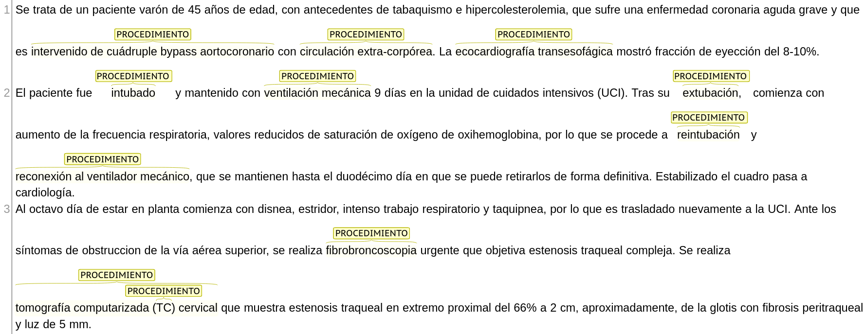
A 45-year-old male patient with a history of smoking and hypercholesterolemia, suffering from severe acute coronary artery disease, underwent quadruple coronary artery bypass grafting with extracoronary circulation. Transesophageal echocardiography showed an ejection fraction of 8-10%.
The patient was intubated and kept on mechanical ventilation for 9 days in the intensive care unit (ICU). After extubation, he began to experience increased respiratory frequency, reduced oxygen saturation and oxyhaemoglobin values, so he was reintubated and reconnected to the mechanical ventilator, which was maintained until the twelfth day, when it could be definitively withdrawn. Once the patient's condition stabilised, he was transferred to the cardiology department.
On the eighth day on the ward he began to experience dyspnoea, stridor, intense respiratory work and tachypnoea, for which he was transferred back to the ICU. Given the symptoms of upper airway obstruction, an urgent fibrobronchoscopy was performed, which revealed complex tracheal stenosis. A cervical computed tomography (CT) scan showed tracheal stenosis at the proximal end of 66% at approximately 2 cm from the glottis with peritracheal fibrosis and a 5 mm lumen.

(A) Biopsy obtained by videothoracoscopy of the mass showed that it consisted of a proliferation of Langerhans cells with abundant eosinophils, histochemical staining showed positivity for S-100 and CD1a, being negative for lymphoid markers, all these findings were compatible with the diagnosis of LCH. After receiving 2 lines of chemotherapy treatment and without obtaining an evident response, it was necessary to initiate radiotherapy therapy, during which a lymphadenopathy appeared in the supraclavicular location, which after being biopsied showed evidence of nodular sclerosis-type Hodgkin's disease associated with LCH; after no response to conventional treatment was observed, autotransplantation of peripheral blood haematopoietic precursors was carried out, achieving complete remission of both diseases to date.

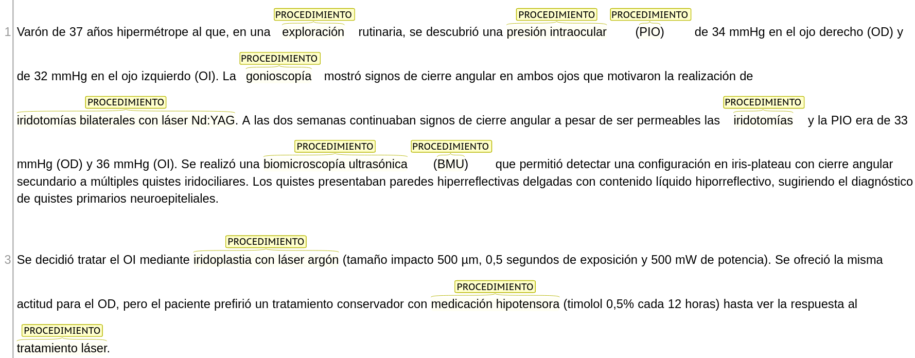
37-year-old hyperopic male who, on routine examination, was found to have an intraocular pressure (IOP) of 34 mmHg in the right eye (RE) and 32 mmHg in the left eye (LE). Gonioscopy showed signs of angular closure in both eyes leading to bilateral Nd:YAG laser iridotomies. At two weeks signs of angular closure continued despite the iridotomies being patent and the IOP was 33 mmHg (RE) and 36 mmHg (LE). Ultrasonic biomicroscopy (UBB) was performed and detected an iris-plateau configuration with angular closure secondary to multiple iridociliary cysts. The cysts had thin hyperreflective walls with hyporeflective fluid content, suggesting the diagnosis of primary neuroepithelial cysts.
It was decided to treat the LE by argon laser iridoplasty (impact size 500 µm, 0.5 seconds exposure and 500 mW power). The same approach was offered for the RE, but the patient preferred conservative treatment with hypotensive medication (timolol 0.5% every 12 hours) until the response to the laser treatment was seen.
Normalization
All entities in the corpus were normalized to SNOMED CT concepts.


